Oxford University Museum 2011
The U3A geology group visited the Oxford University Museum of Natural History on Wednesday 18 May 2011.
Click here for
Visit report
A group of about 30 Malvern U3A Geology Group Members arrived in Oxford with varying degrees of expectation at what they were to experience during their visit to the Geological section of the OUMNH.
The Museum houses the University’s collection of zoological, entomological and geological specimens. The building is Grade 1 listed and is a delight in itself. Even the columns and cornices are named examples of different rocks and samples.
We split into two groups and were first given a tour of the public section with an emphasis on the Geology exhibits. The presentation, simplicity and layout are outstanding. The Museum caters for all levels of interest and knowledge from school children to geology enthusiasts.
The Museum holds approximately 500,000 specimens of which 350,000 have been catalogued. Of particular interest to us were exhibits from Dr Ralph Grindrod, one of the early Malvern Water Cure practioners, whose geology collection had been purchased by the Museum.
We had the privilege of going behind the scenes to see where the majority of the specimens are stored after being catalogued. We saw in particular specimens from the Silurian era, largely from the Welsh Basin, containing many well preserved fossils. Their excellent quality was probably the result of being buried in volcanic ash and bentonite clay which has protected and preserved them from crushing.
We also saw an amazing demonstration of computer technology which creates detailed 3D images of these fossils by taking hundreds of cross sections, a process involving great patience as it can take as long as six months to complete.
OUMNH is truly an outstanding Museum and our visit was enhanced by the opportunity to go behind the scenes to see not only unique specimens but also current research work in action.
Pictures

The first group are given guided tour around the museum exhibits.

A view of the museum. Note the first group in the middle centre.
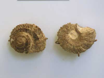
Local fossils #1
Two
gastropod from the display of Silurian fossils in the main part of the Museum.
There are Poleumita rugosa from Ledbury.
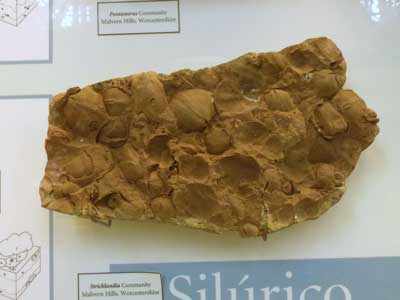
Local fossils #2
A community of Silurian Stricklandia brachiopod fossils from the Malvern Hills.
Again, the main part of the Museum.
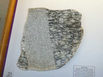
From a display of interesting rocks, in the main part of the Museum.
The
large white eye-shaped crystals of feldspar are called augen, from the German word for eye.
The rock has been stretched, or sheared, around them, producing the wavy
texture.
The vein of even-textured granite was a later igneous intrusion.

The second part of the tour, we go “behind the scenes” to see the work and exhibits that are normally hidden from the public.
The remaining pictures are from that part of the tour.

Until fairly recently, it was thought that there were no fossils earlier than Cambrian times.
A frond like fossil was then found by a school boy in Charnwood Forest, which is believed to be Ediacaran in age.

A Devonian fish, Cheirolepis.

We were introduced to a novel technique for generating 3-D images of fossils.
The fossils are “preserved” in nodules, which has the effect of preserving the soft tissues, albeit in an altered form.
Many very thin sections are then removed from the sample, and scanned into a computer database.
Software can then reconstruct the 3-D images from this data. This process is called tomography.
The pictures below shows a nodule, and the results of producing the thin sections.
For an in-depth explanation on the process, written by the Royal Society, click here.

The results, viewing a soft bodied organism on the computer screen.